The unsettling tumor resembled a malformed human body, complete with organs and limbs.
Disturbing images of an exceptionally rare tumor that developed inside a woman’s ovary have resurfaced online, leaving many stunned.
More than two decades ago, a 25-year-old Japanese woman underwent surgery to remove a tumor discovered in her reproductive system. However, what surgeons found during the procedure was far from ordinary—it was horrifying.
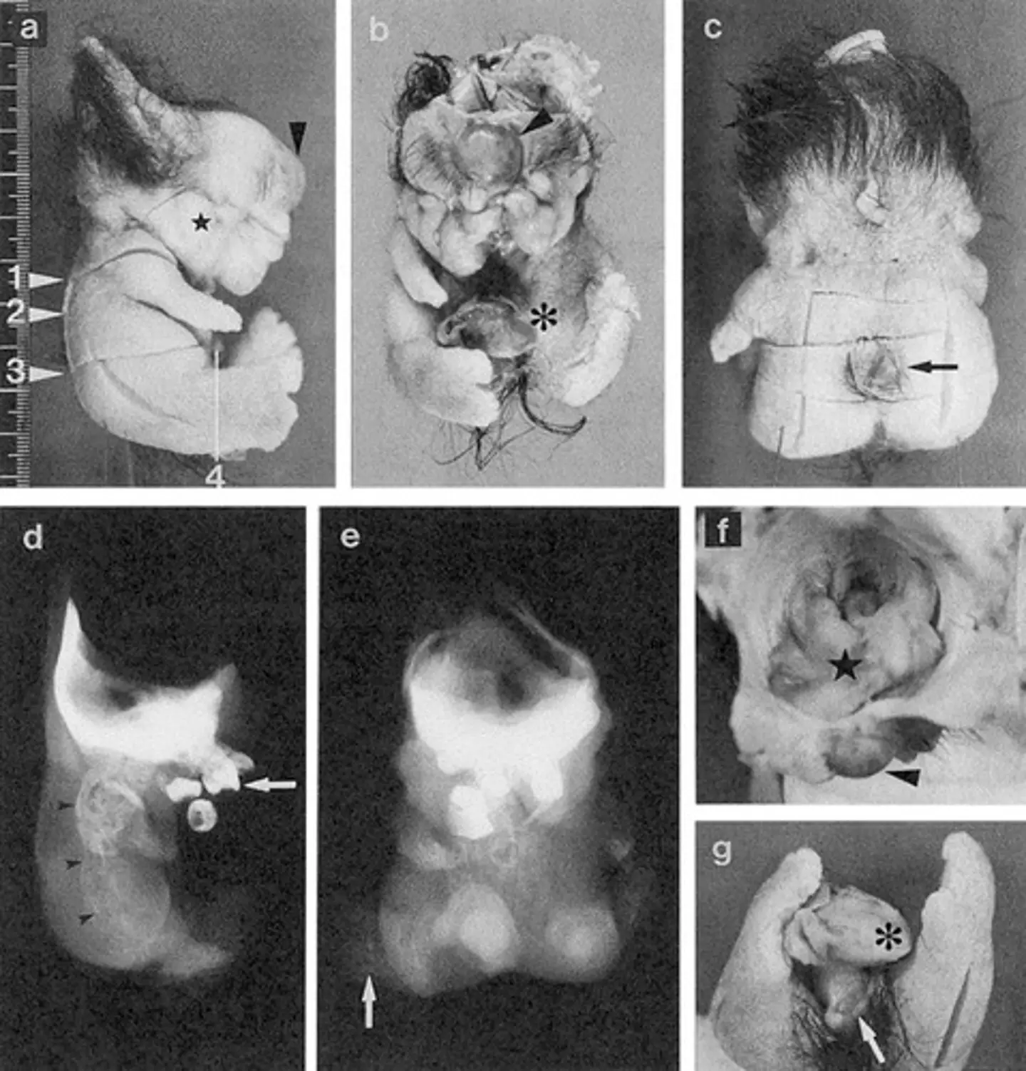
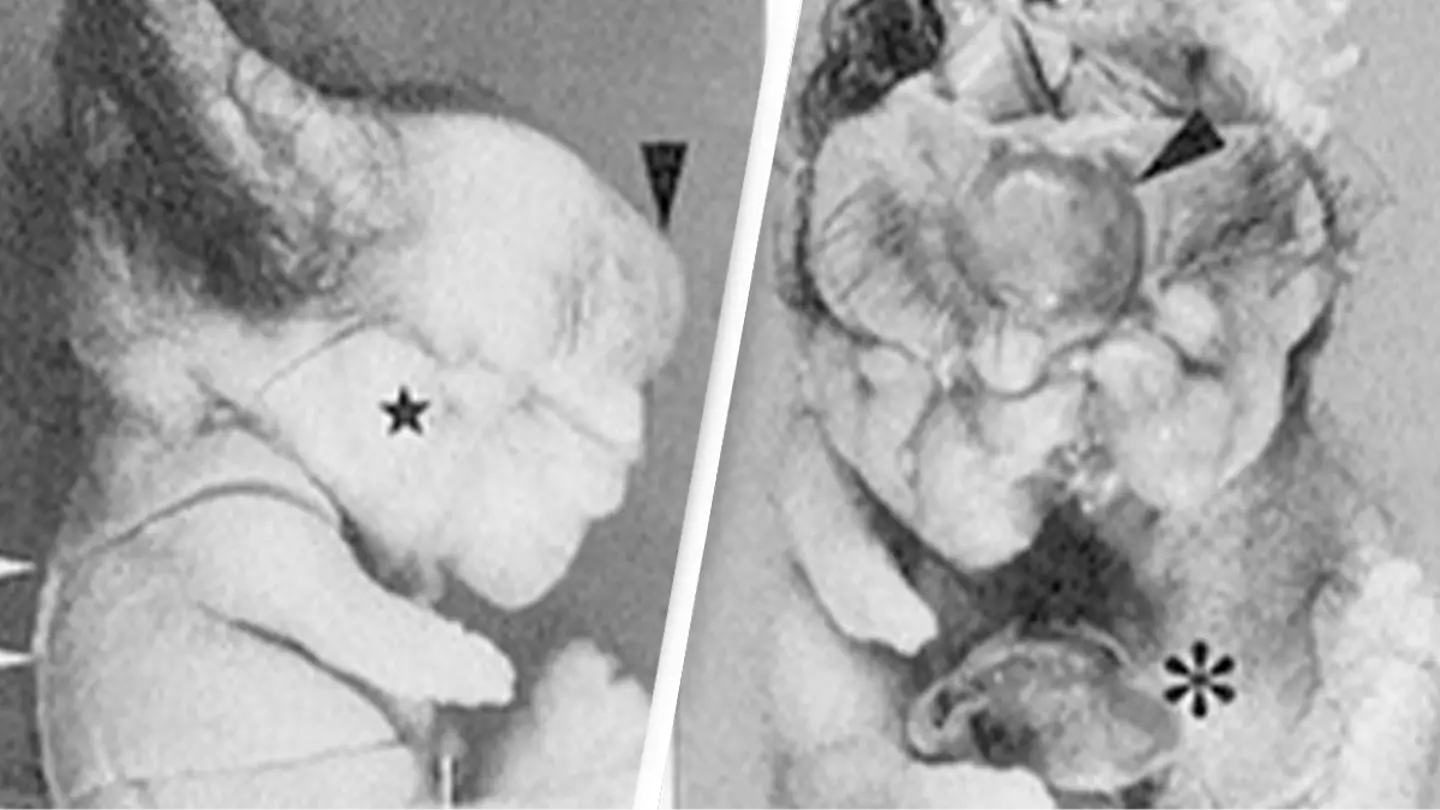
The tumor was unlike typical ovarian growths, as it had developed human-like features, including an eye, an ear, and even a brain.
The Shocking Discovery
Doctors initially diagnosed the tumor as a mature teratoma, a type of benign ovarian tumor that contains tissues from different layers of the body—such as hair, teeth, or skin. While teratomas are not uncommon, this particular case was highly unusual.
A study detailing the case was published in the National Library of Medicine in 2004. Researchers noted that the tumor demonstrated “considerable differentiation,” meaning its growth mimicked the development of an actual human body.
The tumor, described as having a “doll-like” appearance, featured limbs, teeth, and hair, as well as a more organized structure.

A Rare Type of Tumor: Fetiform Teratoma
Doctors ultimately classified the growth as a mature fetiform teratoma (homunculus). According to Cleveland Clinic, this type of dermoid cyst contains living tissues and often resembles a malformed fetus.
The tumor’s internal structure was astonishing. Researchers identified a wide range of components, including:
- Brain tissue
- An eye
- Spinal nerves and bones
- An ear
- Teeth
- Thyroid gland
- Gut
- Blood vessels
- Phallic cavernous tissue
The study’s abstract revealed: “A solid mass within the tumor was found to have a head, trunk, and extremities.”
Detailed Body Organization
What made this tumor even more unsettling was its detailed organization. The researchers described it as having a clear anterior-posterior (front-back), ventral-dorsal (top-bottom), and left-right orientation. The organs were spatially well-arranged, similar to a developing human body.
For instance, the eye was positioned at the front of the head, the thyroid gland sat in front of the trachea, and the gut was located deep within the trunk. This eerie level of development suggested that the tumor followed biological rules similar to those seen in normal human development.

The researchers proposed that the ability to organize a body plan may be “conserved and transmitted,” even in cases of parthenogenesis—a form of asexual reproduction.
A Rare Phenomenon with Valuable Insights
Although mature cystic teratomas are usually benign and don’t receive much attention, the researchers emphasized the importance of studying such tumors.
“Precise analyses of such tumors may significantly enhance our understanding of both parthenogenetic and normal human development,” they wrote.
The authors of the study were Naohiko Kuno, Kenji Kadomatsu, Makoto Nakamura, Takahiko Miwa-Fukuchi, Norio Hirabayashi, and Takao Ishizuka.
Similar Cases in the Past
This isn’t the only known case of a highly developed teratoma. A similar tumor was discovered in a 16-year-old Japanese girl during surgery to remove her appendix. That tumor also contained human-like tissues, further highlighting the fascinating and unsettling nature of teratomas.
Such cases, though incredibly rare, offer valuable insights into human biology and development. However, they also serve as a stark reminder of the complex and sometimes baffling mysteries of the human body.

